Strategizing from 7 cities across the globe
The Erasure of Traumatic Memories through CRISPR Gene Editing of Engram Neurons in the Hippocampus
Due to the advancements in the scientific field, scientists are now able to edit the memory’s molecular basis directly. Recent inventions such as CRISPR-Cas9 are able to modify particular genes in cells with extreme accuracy. Moreover, adeno-associated viral (AAV) vectors make it possible to target this modification to specific neurons, such as the engram cells in the hippocampus, and even regulate the timing of this modification with the use of an antibiotic called doxycycline (Dox) (Wang et al., 2019). Thus, this research proposal will investigate the hypothesis that CRISPR-Cas9 gene editing can be used to disrupt traumatic memories by targeting both the engram cells and their synaptic connections in the hippocampus, because memory is encoded by specific neurons that form the engram and by the strengthened synapses between them. Therefore, using inducible AAV vectors to deliver CRISPR systems against plasticity genes like CREB and Arc holds great potential in disrupting these traumatic memories, freeing patients from PTSD and similar disorders. This research will synthesize evidence on targeting both engram cells and synaptic plasticity, examine the feasibility of using advanced AAV vectors, and thoroughly analyze the ethical implications of such a direct intervention into the human memory.
ROOTSHEALTHCAREMENTAL HEALTHNEUROSCIENCETECHNOLOGYGENETICSPSYCHIATRY
Cindy Aridi
5/23/202611 min read

Introduction and Background
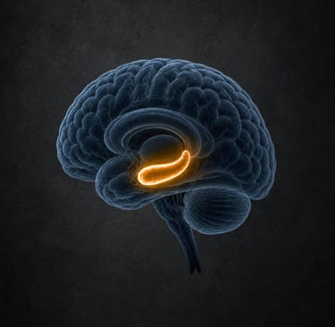
The only way by which an individual is able to recall the past and grow through experiences and mistakes is through memory. In the past, memory was just an abstract idea in the field of Psychology. However, as science became more developed, it was discovered that memories are permanent physical or chemical changes in the neural tissues. They are due to the activation of networks of engram cells that are found in existing neural tissues, where each network is responsible for a specific memory (Josselyn & Tonegawa, 2020).These engram cells are initially activated during learning and then reactivated when the memory is recalled.
Long-term memory is a process that originates from synaptic plasticity. During the formation of a memory, the process of long-term potentiation (LTP) occurs where the synapses between the engram cells increase remarkably in strength. This process is supported by the expression of some genes such as CREB (cAMP response element-binding protein), which is responsible for the initiation of the genetic pathways in long-term potentiation, and Arc (Activity-regulated cytoskeleton-associated protein), which is crucial for the stabilization of the synaptic interactions responsible for preserving the memory (Kandel, Dudai, & Mayford, 2014).
After some time, benign memories may no longer be recalled anymore, and eventually, they may fade away. However, this is not the case with traumatic memories. Traumatic memories are situations where the traumatized individual experiences extreme fear and vulnerability (American Psychiatric Association, 2013). Due to the strong influence these traumatic memories have on one’s psychological state, there is a high probability that they will be recalled from time to time even involuntarily, causing the victim to relive the psychological distress and horror again.
This distress caused by the involuntary re-experiencing of traumatic memories often leads to several mental illnesses such as post-traumatic stress disorder (PTSD). These types of disorders affect millions of people worldwide and obstruct daily functioning. There are current treatments for patients with PTSD, such as prolonged exposure and cognitive behavioral therapy (CBT). The way these treatments work is by breaking the bond between recalling the traumatic memory and suffering afterward from psychological distress, and that is through presenting the traumatic experience in a safe and non-threatening environment. In this way, the patient will still recall the memory, but will tackle it with a calmer and healthier reaction (Watkins et al., 2018). However, as previous research has shown, only 49% to 70% of the patients benefit from the first phase of treatment, and relapse rates remain high (Watkins et al., 2018). This result is due to the fact that the traumatic memories are still present, and these treatments target the symptoms of PTSD rather than the root cause, as the engrams are already activated.
Due to the advancements in the scientific field, scientists are now able to edit the memory’s molecular basis directly. Recent inventions such as CRISPR-Cas9 are able to modify particular genes in cells with extreme accuracy. Moreover, adeno-associated viral (AAV) vectors make it possible to target this modification to specific neurons, such as the engram cells in the hippocampus, and even regulate the timing of this modification with the use of an antibiotic called doxycycline (Dox) (Wang et al., 2019).
Thus, this research proposal will investigate the hypothesis that CRISPR-Cas9 gene editing can be used to disrupt traumatic memories by targeting both the engram cells and their synaptic connections in the hippocampus, because memory is encoded by specific neurons that form the engram and by the strengthened synapses between them. Therefore, using inducible AAV vectors to deliver CRISPR systems against plasticity genes like CREB and Arc holds great potential in disrupting these traumatic memories, freeing patients from PTSD and similar disorders. This research will synthesize evidence on targeting both engram cells and synaptic plasticity, examine the feasibility of using advanced AAV vectors, and thoroughly analyze the ethical implications of such a direct intervention into the human memory.
Targeting Engram Cells
Richard Wolfgang Semon was the first scientist to come up with the idea of “engram cells”. He proposed that an experience in one’s life causes the activation of a group of cells causing them to undergo permanent physical and/or chemical changes to retain the information (Josselyn & Tonegawa, 2020). He claimed that in order to recall the memory, this group of cells has to be reactivated (Josselyn & Tonegawa, 2020). For several decades, this proposal stayed purely theoretical. This is evident as Karl Lashley, a geneticist and psychologist, attempted to locate engram cells for 30 years but still failed to do so (Josselyn & Tonegawa, 2020). However, significant technological advancements in the field of genetics transformed this theory into action. For example, loss-of-function experiments proved that eliminating these particular engram cells impairs memory retrieval, and gain-of-function experiments showed that even without any natural sensory signal that might remind the person of the experience, artificially reactivating these engram cells via optogenetics leads to memory recall (Josselyn & Tonegawa, 2020). Thus, the presence of these engram cells provides a tangible target for therapeutic interventions related to memory loss and retrieval. This presents a highly promising solution for erasing traumatic memories.
A significant example of the loss-of-function experiment is the study conducted by Josselyn and colleagues in 2009 (Josselyn & Tonegawa, 2020). The researchers conducted an experiment on mice where they targeted the engram cells in the lateral amygdala, the brain region responsible for auditory fear memory. First, they injected a viral vector to induce the overexpression of the CREB transcription factor in a random group of neurons in the lateral amygdala. Only the neurons that were selected for the CREB injection were overly excitable and more ready to be incorporated into the fear memory formation process (Josselyn & Tonegawa, 2020). Then, they exposed the mice to a particular neutral sound; however, after every play, they ended it with a foot shock, which is an electrical shock in the feet that causes extreme fear and automatic reactions such as freezing and jumping (Josselyn & Tonegawa, 2020). This was done to let the mice learn the pattern and associate the sound with fear. Thus, after the memory was formed, the researchers utilized an enzyme that causes apoptosis, caspase-9, to eliminate all the engram cells that were overexpressed by CREB (Josselyn & Tonegawa, 2020). As a result, after the mice were exposed to the same sound again, they did not show a freezing reaction (Josselyn & Tonegawa, 2020). This shows the complete deletion of the auditory fear memory in the mice. Moreover, this experiment was proven to be highly specific since the lateral amygdala was not damaged and the mice could learn other fear memories easily. Moreover, the elimination of the neurons that were not selected for CREB overexpression had no effect on memory recall (Josselyn & Tonegawa, 2020). Even though this experiment was done on mice and not humans, it stands as significant evidence that elimination of the specific engram cells leads to the erasure of the associated memory.
Targeting Synapses
Even though targeting the engram cells has been proven to be efficient in fear-memory erasure, this method holds hidden risks that include deleting other unrelated memories that are controlled by the same group of engram cells. A single engram cell has the ability to participate in the formation of many memories; thus, its elimination causes the deletion of all of these memories, leading to several gaps in one’s memory and a significant threat to one’s personal narrative (Josselyn & Tonegawa, 2020). This limitation encourages scientists to try another method, which is targeting the synapses between the engram cells rather than the cells themselves. The “synaptic plasticity hypothesis” posits that the complete formation of a memory involves both the engram cells and the synapses between them (Kandel et al., 2014). These synapses are responsible for the process of long-term potentiation (LTP). By targeting the strengthened synapses between the engram cells, the memory itself would still be present; however, the association of the memory with the fear response would be broken.
A prominent experiment conducted by Nabavi and colleagues in 2014 presents a case in which LTP is reversed to Long-Term Depression (LTD) and shows its effect on fear memory erasure. First, the researchers injected a genetically modified virus that contains a gene encoding the light-sensitive protein Channelrhodopsin-2 (ChR2) into the lateral amygdala of a group of rats. This gene was controlled by a particular promoter (c-Fos) which becomes activated in highly active neurons only (Nabavi et al., 2014). Then, they exposed this group of rats to a “fear conditioning paradigm”, which is basically associating a neutral cue such as a tone with an electric shock such as the foot shock so that later on, the cue alone will trigger the fear response. When the rats learned the fear memory, only the specific engram cells that were responsible for forming the memory-produced ChR2 (Nabavi et al., 2014). After blue light was introduced to activate these targeted engram cells, this optogenetic activation induced the freezing response in the rats, confirming the presence of the memory in these cells. In order to erase this memory, the researchers applied the LTD protocol. Instead of briefly exposing the neurons to blue light to activate them, they utilized a series of light pulses delivered at a slow rate to induce artificial LTD (Nabavi et al., 2014). This protocol removes the AMPA receptors on the strong synapses by endocytosis, making the synaptic connection weaker. After the process is completed, the rats do not show any signs of fear as they are exposed to the original tone. This proves that the fear memory is erased in the rats. However, a limitation to this process is the possibility to retrieve the strengthened synapses. For instance, in the same experiment, after they exposed the neurons again to a series of high frequency light pulses, the synapses established Long-Term Potentiation and the memory was restored (Nabavi et al., 2014). This approach does not promise the permanent erasure of the association between the memory and the fear response. There remains a probability that this association may recover over time since a single intervention might not be enough to permanently erase the fear response.
Another significant study concerning the long-term erasure of a memory was done by Shema and other colleagues in 2007. First, the researchers placed a group of rats in a “taste-aversion paradigm”, where they were given a new sweet flavor which was saccharin, and were then injected with Lithium Chloride to induce nausea (Shema et al., 2007). Thus, these rats associated the sweet taste with feeling nauseous. It was proven that after this experiment, these rats avoided drinking anything sweet. Because taste memories are stored in the insular cortex, the researchers injected a compound named Zeta Inhibitory Peptide (ZIP) in this brain region after the full consolidation of the memory. ZIP is an inhibitor of the PKMzeta protein which is responsible for maintaining the strengthened synapses (Shema et al., 2007). After a few days, the researchers gave the rats the same sweet taste and the rats drank it normally as if they had never associated it with feeling nauseous. The control group of rats avoided drinking it. This proves the erasure of this memory in the experimental group. In order to prove that this experiment did not damage the whole brain, the researchers successfully introduced a new taste memory, and the rats were fully able to learn the new association (Shema et al., 2007). Although this method provides a more permanent and stable erasure of memories, a new limitation is present due to the injection of ZIP in all the insula cortex. This lack of specificity might lead to the erasure of many memories present in that region and supported by the PKMzeta protein. This presents a huge threat to the human memory and the individual’s narrative.
AAV Vectors
Adeno-associated virus (AAV) vectors are an advanced and powerful invention in the field of genetics and neuroscience research. They are ideal for interventions in the brain regions due to their high specificity, efficiency, and safety. In studying chronic neurological diseases, AAV vectors are benign and they cause only a minimal immune response. Moreover, they have the ability to cause long-term gene expression in neurons that are not dividing (Wang et al., 2019).
Unlike retroviruses, AAV vectors insert their genetic material, a single-stranded DNA, as separate genetic material, without integrating it into the host’s chromosomes, a process that might lead to mutations. This causes stable gene expression without disturbing the host’s original genetic material.
Their extreme specificity and flexibility make them even more promising for the future. There are different AAV serotypes specific to different brain regions. For instance, AAV9 is utilized in direct injections in the CNS region (Chan et al., 2017). Meanwhile, when AAV-PHP.eB is injected into the blood, it has the ability to cross the blood-brain barrier and allow widespread delivery across the brain (Chan et al., 2017). Moreover, researchers are also able to specify exactly which cells should express the transferred gene through the use of promoters such as c-Fos or Arc. These promoters often work on active neurons, such as the ones that become activated during remembering a fear memory (Liu et al., 2012). Inducible systems like the tetracycline-dependent switches (Tet-On/Tet-Off) allow the researchers to choose when the gene expression occurs via the antibiotic doxycycline (Dox) (Reijmers et al., 2007). When the experimental animal gets doxycycline (Dox) through its food or in its drinking water, the expression of the gene remains inactivated. When Dox is withdrawn, the expression is activated, allowing it to function at the desired time (Reijmers et al., 2007).
In the context of human memory modification, the AAV vector that should be used will deliver the genetic instructions rather than the gene itself. It will carry the DNA sequence that encodes the Cas9 enzyme (which cuts DNA) and the guide RNAs (gRNAs) that send the Cas9 enzyme to the two genes that are responsible for memory formation, Arc and Creb (Liu et al., 2012). Thus, the mechanism is as follows. First, the AAV vector enters the genes responsible for synaptic plasticity, but it stays dormant. It is important to note that when the memory is formed, Dox is given to the experimental animal. Then, when the memory is recalled, Dox will be removed. In this case, only the neurons that become active in the process will express the c-Fos promoter. When the necessary promoter is active and Dox is removed, the Cas9 gene and the gRNAs will only be expressed in the engram neurons. This will disrupt the Arc and Creb genes and either weaken or erase the recalled memory.
Conclusion
AAV vectors with CRISPR technology hold great potential for targeting specific neurons responsible for traumatic memories. Because of their specificity and precision, they make selectively targeting and disrupting genes in the engram cells possible, allowing researchers to erase the traumatic memories without disrupting other processes. However, this method of manipulating memories raises important ethical concerns. The future of this method is still not clear, even in animal experiments. Thus, there might be some unforeseen consequences that could be threatening in a certain way. Moreover, if memories can be manipulated, this will definitely raise questions about identity, consent, and autonomy. Therefore, there should be strict and clear ethical frameworks for this method, avoiding any misuse and minimizing harmful consequences.
DOWNLOAD THE FULL DOCUMENT
References:
American Psychiatric Association. (2013). Diagnostic and statistical manual of mental disorders (5th ed.). https://doi.org/10.1176/appi.books.9780890425596
Chan, K. Y., Jang, M. J., Yoo, B. B., Greenbaum, A., Ravi, N., Wu, W.-L., Sánchez-Guardado, L., Lois, C., Mazmanian, S. K., Deverman, B. E., & Gradinaru, V. (2017). Engineered AAVs for efficient noninvasive gene delivery to the central and peripheral nervous systems. PubMed. PMID: 28671695. https://pubmed.ncbi.nlm.nih.gov/28671695/
Josselyn, S. A., & Tonegawa, S. (2020). Memory engrams: Recalling the past and imagining the future. Science, 367(6473), eaaw4325. https://doi.org/10.1126/science.aaw4325
Liu, X., Ramirez, S., Pang, P. T., Puryear, C. B., Govindarajan, A., Deisseroth, K., & Tonegawa, S. (2012). Optogenetic stimulation of a hippocampal engram activates fear memory recall. PubMed. PMID: 22441246. https://pubmed.ncbi.nlm.nih.gov/22441246/
Nabavi, S., Fox, R., Proulx, C. D., Lin, J. Y., Tsien, R. Y., & Malinow, R. (2014). Engineering a memory with LTD and LTP. Nature, 511(7509), 348–352. https://doi.org/10.1038/nature13294
Reijmers, L. G., Perkins, B. L., Matsuo, N., & Mayford, M. (2007). Localization of a stable neural correlate of associative memory. Science, 317(5842), 1230–1233. https://doi.org/10.1126/science.1143839
Shema, R., Sacktor, T. C., & Dudai, Y. (2007). Rapid erasure of long-term memory associations in the cortex by an inhibitor of PKMζ. Science, 317(5840), 951–953. https://doi.org/10.1126/science.1144334
Wang, D., Tai, P. W. L., & Gao, G. (2019). Adeno-associated virus vector as a platform for gene therapy delivery. PubMed. PMID: 30710128. https://pubmed.ncbi.nlm.nih.gov/30710128/
Watkins, L. E., Sprang, K. R., & Rothbaum, B. O. (2018). Treating PTSD: A review of evidence-based psychotherapy interventions. Frontiers in Behavioral Neuroscience, 12, 258. https://doi.org/10.3389/fnbeh.2018.00258